DOI: 10.25504/FAIRsharing.9f9f9c
Repository

3AMY
The structure shown is the crystal structure of human CK2α complexed with apigenin (PDB 3AMY)
Image rendered by: Amidhaar Bhardwaj
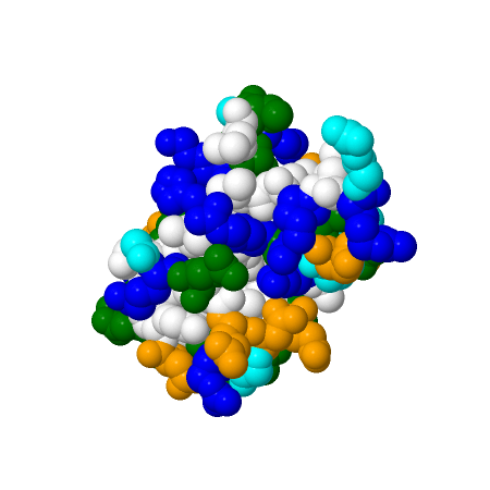
1UBQ
The structure shown is the crystal structure of ubiquitin refined at 1.8 Å resolution (PDB 1UBQ), a 76-residue regulatory protein from human erythrocytes (Homo sapiens).
Image rendered by: Amidhaar Bhardwaj
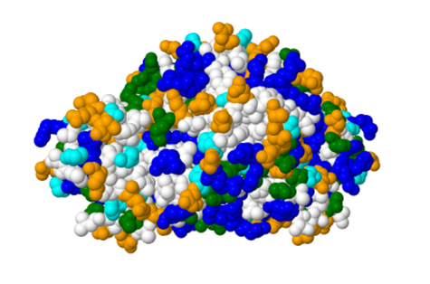
3CHI
The structure shown is the crystal structure of di-iron AurF in its monoclinic form (PDB 3CHI), a p-aminobenzoate N-oxygenase from Streptomyces thioluteus.
Image rendered by: Karen Martinez
Loading...